Cubital Tunnel Syndrome

By Marianne Pilgrim, OTR/L, CHT

Cubital TunnelMost people are familiar with carpal tunnel syndrome, but few know about a similar condition: cubital tunnel syndrome. While carpal tunnel syndrome affects the median nerve at the wrist, cubital tunnel syndrome affects the ulnar nerve at the elbow.  This is the same nerve that is irritated when you hit your “funny bone” and you feel a tingling sensation running down to your fingers.

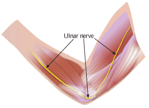
The ulnar nerve originates from the spinal cord at the neck, and it runs through the entire arm. This nerve provides sensation to the small and ring fingers and strength to the small muscles of the hand.  The ulnar nerve may be compressed as it passes behind the medial epicondyle, a bony bump on the inside of the elbow.  Cubital tunnel syndrome is a pathologic compression, friction or traction of the ulnar nerve in the elbow region.  Symptoms may include impaired sensation of the small and ring fingers, burning pain, weak grip and clumsiness of the hand.

Common causes of cubital tunnel syndrome include direct pressure, prolonged bending of the elbow, repetitive movement, congenital anomolies, inflammation and bony changes. Conservative treatment of this condition may include use of NSAIDs (non-steroidal anti-inflammatory drugs), education by a therapist regarding proper ergonomics and techniques to avoid aggravating positions or activities, and nighttime splinting or daytime padding of the elbow.  If the symptoms do not respond to conservative treatment, surgery may be required to provide more space for the ulnar nerve. In a procedure known as ulnar nerve transposition, the nerve is moved to the front of the elbow.  Therapy is usually prescribed after surgery to increase range of motion, increase strength and provide scar management.

Hip Pain – Causes and Treatment

By Lisa Gemmel, MPT

hippainThe hip is a ball and socket joint between the femur (thigh bone) and the pelvis. The head of the femur fits into the acetabulum (hip socket). The acetabular labrum, a ring of cartilage inside the joint, acts like a suction cup to keep the joint together. The hip joint capsule contains muscles and tendons that act upon the joint.  It also has two bursas, fluid filled sacs that prevent friction between bone and soft tissue.  The bursa on the outer part of the hip prevents friction between the head of the femur and a band of connective tissue called the iliotibial band.  The other bursa is located in the front of the hip, under the iliopsoas muscle. Any of these structures, as well as the lumbar spine, can cause hip pain.

Some common diagnoses associated with hip pain include osteoarthritis, labral tear, bursitis, tendonitis, iliotibial band syndrome, and muscle strains.  These conditions can be caused by any of the following:  trauma, degeneration, overuse, and structural abnormalities such as leg length discrepancy. Once the proper diagnosis is made, treatment can be initiated.  Treatment can include anti-inflammatory medicines, cortisone injections and physical therapy.  Physical therapy includes therapeutic exercise, modalities for pain and inflammation, soft tissue or joint mobilizations, and instruction in correct movement patterns.

Hamstring Injuries: Causes and Treatments

By Nancy Simpkins

A dancer’s arabesque, a baseball player’s sprint for first base, and a football player’s place kick are all activities that depend on healthy hamstrings. The hamstrings are three muscles at the back of the thigh: the semitendinosus, semimembranosus, and biceps femoris. They start at the ischial tuberosity, (also called the sitz bone) at the back of the pelvis, and are connected to the lower leg by the hamstring tendons.

The term “hamstrung” acquired the meaning of “powerless” or “crippled” from the historical practice of cutting domestic servants’ hamstring tendons so they couldn’t escape. Now, hamstring injuries are most prevalent among athletes, particularly those who run, jump or kick.

The hamstrings help bend the knee and extend the hip, and they work in opposition to the quadriceps, the muscles on the front of the thigh. The quadriceps are normally stronger than the hamstrings, but if they become too much stronger, the resulting muscular imbalance puts the hamstrings at risk of injury. Tight hamstrings are also prone to injury.

What Causes Hamstring Injuries?

Hamstring strains can range from a partial tear of the muscle to a complete rupture. Symptoms range from tightness and pain in the back of the thigh to inability to walk or straighten the knee. There may be bruising in the back of the thigh, swelling, or muscle spasms. Sudden movement of the knee can cause the hamstring tendon to rupture. Symptoms include sudden pain at the back of the knee followed by swelling, and pain when bending the knee against resistance.

How Are Hamstring Injuries Treated?

Treatment of hamstring injuries usually starts with rest, ice, compression and elevation. Physical therapy may be recommended, and will include range of motion and strengthening exercises. Some doctors try to stimulate faster healing by injecting platelets of the patient’s own blood into the site of the injury, a technique called platelet rich plasma therapy (PRP). Severe hamstring injuries may require surgery.

To avoid hamstring injuries, it is important to warm up and stretch before exercise, and to do strengthening exercises as well. Athletes who have suffered one hamstring injury are at increased risk for another one. A physical therapist can teach you how to keep your hamstrings flexible and strong.

Baker’s Cysts

bakerscyst2A Baker’s cyst, also called a popliteal cyst, is a fluid-filled lump behind the knee. It is named after William Morrant Baker, the doctor who first described it. The cyst occurs when the popliteal bursa, a soft pouch behind the knee, accumulates excess fluid and expands. It may feel like a water-filled balloon.

The fluid in a Baker’s cyst is synovial fluid, which normally lubricates the joint. Excess fluid can build up after an injury such as a tear of the meniscal cartilage in the knee. These cysts are also associated with knee arthritis.

Symptoms may include stiffness, pain, swelling, or a decrease in range of motion. A cyst that ruptures may also cause bruising on the back of the knee and calf. Sometimes a blood clot can be confused with a ruptured Baker’s cyst, but in the case of a blood clot, immediate treatment is required. If your calf is red and swollen, you should seek medical attention.

Baker’s cysts may go away without treatment, but large and painful cysts are usually treated. If you have a Baker’s cyst, a physical therapist can teach you range of motion and strengthening exercises to reduce symptoms and maintain knee function. Your therapist may also apply ice or a compression bandage to reduce swelling. Your doctor may also reduce swelling with a corticosteroid injection or by draining the fluid from the cyst. If the cyst is caused by torn cartilage or arthritis, surgery may be recommended.

Osteoarthritis of the Hand

osteoarthritisBy Marianne Pilgrim, OTR/L, CHT

As summer approaches, many people will resume a favorite pastime: gardening. Engaging in a repetitive activity like gardening can sometimes lead to arthritis, but educating yourself about how to prevent and manage this disease can preserve your joints for many more gardening seasons.

According to the Arthritis Foundation, osteoarthritis, the most common type of arthritis in the hands, affects nearly 27 million Americans.  Osteoarthritis is a degenerative condition in which the cartilage that cushions the bone surfaces at the joints wears down, resulting in pain and stiffness.  The exact cause of osteoarthritis is not known, but joint injury, repetitive overuse, lack of physical activity and heredity are associated with an increased risk of developing the disease.

In the hand, osteoarthritis most commonly develops at the base of the thumb and in the two smaller joints of each finger. Symptoms may include pain, stiffness, swelling, bony nodules and weakness.  Osteoarthritis is diagnosed with a thorough medical history, examination and X-ray.

If you have osteoarthritis, hand therapy can help preserve your joints.   Certified hand therapists educate patients on joint protection techniques, activity modification, stretching and strengthening. They can also fabricate custom resting splints and recommend ergonomic tools for gardening and everyday tasks.  Patients with advanced osteoarthritis that has caused joint damage and limitations in joint function may benefit from surgery.

Shin Splints

By Lisa Gemmel, MPT

shinsplintsShin splints is a disorder affecting the muscle-tendon unit in the lower leg. Symptoms include pain, soreness or tenderness in the front of the lower leg. The condition is brought on by overuse, and while it can be caused by many activities, the most common one is running. People with low arches have a higher than normal risk of shin splints.

Treatment should begin with stopping the activity causing the pain and trying an alternative form of training, such as non-impact underwater running, using an elliptical machine or cycling.  Ice and anti-inflammatory medications such as ibuprofin can also help.  If these measures are not successful in alleviating the pain, or if the pain gets worse, it’s time to seek medical attention from an orthopedist.

After ruling out other causes of pain in the area, such as a stress fracture or compartment syndrome, the orthopedist will refer you to a physical therapist.  The therapist will evaluate you and identify which mechanical stresses are causing the problem. The affected muscle can then be strengthened and conditioned by performing specific exercises and stretches. Heat applied before exercise increases blood flow and warms up the muscles, and ice is used after exercise to reduce inflammation. Orthotics or a change of footwear may be recommended to help cushion the shin and put your foot in the correct alignment, which reduces the strain on the muscle.

Total Knee Replacement

By Nancy Simpkins

kneereplacementWhen arthritis in the knee causes severe pain or disability that is not improved by conservative treatments, surgery to replace the joint may be recommended. In total knee replacement surgery, damaged cartilage is removed, and a metal or plastic joint is attached to the femur (thigh bone), tibia (shin) and patella (knee cap).

A small number of orthopedic surgeons do quadriceps-sparing minimally invasive total knee replacement, which requires a smaller incision than in traditional surgery, and eliminates the need to cut the quadriceps tendon and muscles, and to dislocate the kneecap. Patients benefit from a shorter hospital stay, shorter recovery time and a smaller scar.

After knee replacement surgery, patients receive physical therapy to learn exercises that strengthen the muscles in the knee and increase the range of motion in the joint. Physical therapy can also help reduce pain and swelling. Some patients may need to relearn good standing and walking techniques. Many patients with knee pain develop a habit of using the hips to stand up from a chair, which increases the risk of falling and could lead to future knee osteoarthritis. After knee replacement surgery eliminates the pain, a physical therapist can train these patients to use their quadriceps muscles instead.

In addition to improving function in the knee, exercise can help control weight, which is important since excess weight can reduce the life of the new joint. When knee replacement began in the 1970’s, the life of the replacement joint was estimated at 10 years. Current replacement joints are expected to last closer to 20 years.

Spinal Stenosis

By Hector Mayo, PT

Spinal stenosis is a narrowing of the spine that is usually caused by osteoarthritis. Less common causes include a herniated disc, stiffening of the ligaments in the spine, spinal tumors, injuries, spinalstenosisPaget’s disease and dwarfism. While it is possible to be born with spinal stenosis, most cases develop after age 50.

Spinal stenosis is most common in the lumbar spine, in the lower back, and occurs less frequently in the cervical spine, in the neck. It only rarely affects the thoracic spine, in the mid-back. When the narrowing spine puts pressure on the spinal cord or nerves that branch out from it, pain or numbness in the back, legs, neck, shoulders or arms can result. With spinal stenosis that causes leg pain, walking can exacerbate the problem, while sitting or bending forward can decrease the pain. Stenosis in the cervical spine can cause problems with balance and coordination, and patients may develop a tendency to fall.

In rare cases, spinal stenosis can compress the cauda equina, the bundle of nerve roots that extends from the end of the lumbar spine. This compression is called cauda equina syndrome, and it may cause severe low back pain, bladder and bowel dysfunction, sexual dysfunction, weakness in the legs, and an inability to feel the areas of the body that would sit in a saddle. This condition should be treated immediately to avoid permanent incontinence or paralysis.

In many cases of spinal stenosis, anti-inflammatory medications and physical therapy can relieve symptoms. In physical therapy, patients learn specific exercises to increase strength and flexibility in the muscles and ligaments that support the spine. This can improve posture and alleviate symptoms. Regular exercise can also help with losing weight, which often reduces pain. Some patients receive epidural steroid injections to reduce inflammation and relieve pain. Surgery may be recommended for patients with severe symptoms that are not helped by other treatments.